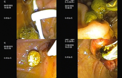
Young 32yr male came with h/o accidental consumption of metallic cap under alcohol influence. Cap was found to be stuck in the upper oesophagus and was removed using rat tooth forceps.
Case : 2
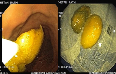
An 18yr boy came with h/o consumption of
mango seeds as challenged by his friend 4 days back. The patient later
developed abdominal pain and vomiting. Endoscopy revealed 2 large mango seeds
almost 5cm each. Both seeds were removed using a snare.
Case : 3
A 2yr kid was brought by parents with h/o coin ingestion. Coin was removed using a roth net.
Case : 4
Upper GI endoscopy in c/o cirrhosis with haematemesis revealed large oesophageal varices, endoscopic variceal band ligation (EVL) was done.
Case : 5
A chronic alcoholic with h/o recent binge came with an episode of haematemesis. Endoscopy revealed Mallory Weiss tear with bleeding vessel. Haemoclip was applied.
Case : 6
A non-alcoholic with h/o ischemic heart disease came with an episode of haematemesis and melena. Endoscopy revealed a gastric ulcer with visible vessel. Haemoclip was applied.
Case : 7
Middle aged woman with h/o dysphagia to solids and liquids for 10 years. Investigations revealed Achalasia Cardia. Balloon dilatation was done to relieve her symptoms.
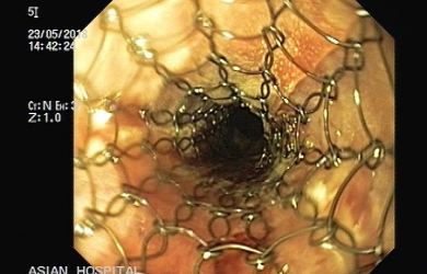
Case : 8
Self expanding oesophageal metallic stenting for carcinoma of oesophagus.
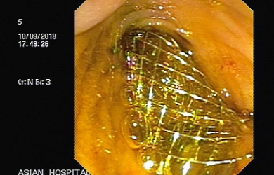
Case : 9
Self expanding oesophageal metallic stenting (stent in stent) for recurrence causing distal obstruction of previously placed SEMS in carcinoma of oesophagus.
Case : 10
Biliary metallic stenting for malignant obstructive jaundice (Inoperable carcinoma head of pancreas)
Case : 11
A 10-year girl with h/o abdominal pain (Acute pancreatitis) followed by recurrent pleural effusion – requiring repeated therapeutic tapping in 1 year was referred to rule out chronic pancreatitis (CP). Previous investigations including EUS did not reveal any changes of CP. Investigations revealed high pleural fluid amylase (>1,000 IU/ml). But, MRCP failed to reveal pancreatic duct (PD) leak. Pancreaticogram during ERCP revealed a duct leak in the tail of the pancreas. PD was stented to relieve the patient’s symptoms and a stent was removed 3 months later. The patient is asymptomatic since then.
Case : 12
Endoscopic Retrograde Cholangio Pancreatography (ERCP) for benign (CBD stones) and malignant (ampullary mass) biliary diseases.